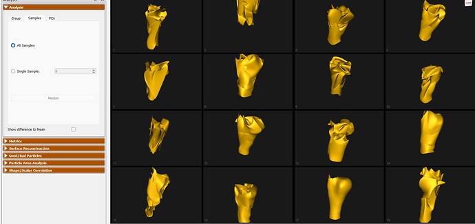
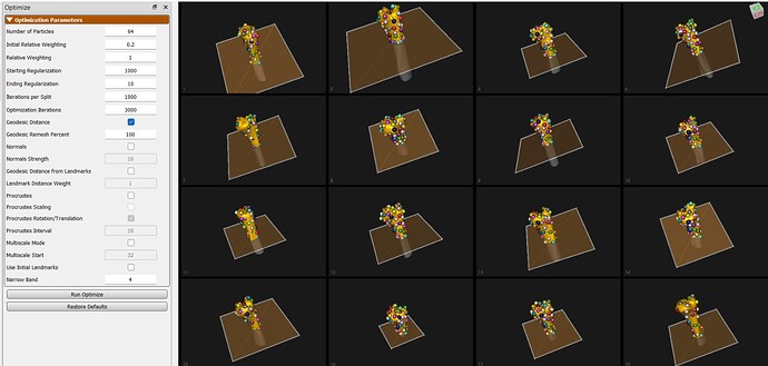
Hi there! I am working on a research project involving ulnar fractures. Specifically, I am trying to identify the fracture type for each patient by analysing 3-D images generated from CT scans. This is my first time using ShapeWorks—thank you for providing such an impressive tool!
After the grooming and optimisation steps in ShapeWorks Studio, the reconstructed bones look inaccurate and the overall alignment is also poor. I am wondering whether I am missing any steps, have mis-configured parameters, or am encountering some other issue in the pipeline.
I have attached the parameter file and several screenshots of my data.
Interestingly, when I add a few initial landmarks the optimisation improves markedly, but I would prefer to avoid manual landmarks if possible.
If anyone with more experience could share advice or point out potential pitfalls, I would greatly appreciate it.
Thank you very much!
There are a few issues here.
- These structures are cut off at different positions. I would recommend setting a consistent cutting plane in Studio as shown here:
- I would recommend increasing the iterations per split to at least 2000 or 3000. They may not be stabilizing well during initialization.
Thank you for your response.
We have followed your recommendations and re-optimized the model.
However, we are still encountering an issue: the resulting modes exhibit spatial translations that we would like to eliminate. We believe this may be due to the alignment being performed on the entire bone, rather than focusing specifically on the yellow region. Additionally, the points on the head of the bone do not align properly across samples.
Do you have any further suggestions that might help us address these issues?
Additionally, we wanted to inquire if there have been any successful cases of ShapeWorks applied to fractured bones. To provide a bit more context, we are attempting to use ShapeWorks software to classify ulnae with fractures, where classification depends on the size of the fracture—in other words, the size of the fragment that separated from the bone. Initially, we tried performing multi-mode optimization including the fractured fragment, but the resulting mean shape and modes were not accurate or useful. Therefore, we decided it was better to proceed only with the bone (excluding the fractured fragment). We are considering the possibility of adding the volume of the detached bone fragment as an additional mode, although we still need to investigate how to obtain this volume using ShapeWorks.
I am attaching images showing the results of the optimization using the planes and metrics you recommended. I have also attached images illustrating how the multi-mode optimization performed, just in case you are curious (I included the optimization and the mean).
Any insights or experiences you could share regarding this type of application would be greatly appreciated.
Thank you very much in advance.
The alignment should be run on the clipped mesh.
Is there a chance you could share a subset of the data with me? amorris@sci.utah.edu


